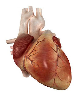
Study on two specially bred strains of mice showed how abnormal addition of the phosphate to a specific heart muscle protein may sabotage the way the protein behaves in a cell, and may damage the way the heart pumps blood around the body. Different people may have more or less altered phosphorylation that might help patients who may benefit from targeted therapies.
A form of heart disease known as heart failure with preserved ejection fraction-the amount of blood squeezed out when the heart contracts impairs the heart’s ability to quickly and efficiently relax between the heart beats and overworking the organ. Common symptoms of heart failure include shortness of breath, but those with the form in which the ejection fraction is preserved at baseline have particular difficulty when they try to increase their activity or exercise.
Heart failure with preserved
ejection fraction does not respond well to common heart failure medications. The condition is common in adults, though some children with genetic disorders of heart muscle proteins share features of this condition. Heart failure was associated with changes in heart muscle cells through altered phosphorylation in the heart muscle protein cardiac troponin I cTnI, which regulates heart contraction.
Researchers examined the function of the mouse hearts through echocardiography as well as measurements with tiny catheters placed in the heart compared to mice without this altered phosphorylation. At baseline the mice with hyperphosphorylation on this specific site experienced a longer time to heart relaxation and lower left ventricular peak filling rate (depressed diastolic function), but the amount of blood ejected during contraction was normal.
The researchers then stimulated both strains of mouse hearts with adrenaline to assess the impact of increased demand on the hearts. The mice with hyperphosphorylation had very limited ability to increase the ejection of blood from the heart compared to the controls in response to adrenaline. However, the mice with hyperphosphorylation did show some improvement in relaxation, though relaxation remained slower than controls at peak drug effect.
Researchers then subject both sets of mice to brief periods of reduced oxygen flow to the heart and then restored the flow of oxygen, they discovered that the hearts of mice with hyperphosphorylation were protected from this form of stress.
haleplushearty.blogspot.com


