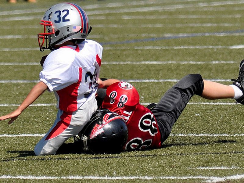
There’s more evidence that football may be changing the brains of adolescent players, and not in a good way.
In a new study, researchers looked at MRI scans of 26 football-playing boys averaging 12 years of age.
Comparing MRIs taken just before the football season and then three months after, the scans revealed that the boys had changes in an important area of the brain called the corpus callosum. That’s a band of nerve fibers that connects and helps “integrate” functions between the two halves of the brain.
The football players’ brain scans were also compared to those taken of a “control” group of 22 boys who didn’t play football.
Clear differences were seen, according to the researchers who presented the findings Thursday at the annual meeting of the Radiological Society of North America (RSNA), in Chicago.
“The years from age 9 to 12 are very important when it comes to brain development,” said lead study author Jeongchul Kim, of Wake Forest School of Medicine in Winston-Salem, N.C.
“The functional regions of the brain are starting to integrate with one another, and players exposed to repetitive brain injuries, even if the amount of impact is small, could be at risk,” he explained in an RSNA news release.
“The body of the corpus callosum is a unique structure that’s somewhat like a bridge connecting the left and right hemispheres of the brain,” Kim said. “When it’s subjected to external forces, some areas will contract and others will expand, just like when a bridge is twisting in the wind.”
So, the new study suggests that repeated hits to the head — as can occur in the rough-and-tumble of football — may be causing the brain changes seen in these young players.
Still, the study is not conclusive, and further research is needed to confirm that repeated blows to the head in football and other youth contact sports could lead to changes in the shape of the corpus callosum during this critical time of brain development, Kim said.
Similar findings were reported on Monday in another study from the RSNA meeting. In that study, researchers at UT Southwestern Medical Center in Dallas compared pre- and post-season MRI scans from 60 football-playing boys aged 9 to 18.
Those researchers saw more gray matter volume in the brains of youngsters who had high-impact hits — but no concussions — over the season.
More gray matter brain tissue indicates that the brain might not be working as well as it could be, explained study author Gowtham Krishnan Murugesan.
One expert who’s seen the effects of brain trauma first hand agreed that young people may not need a full-blown concussion to be neurologically changed by head impacts.
While the sample size in the Wake Forest study is small, “it demonstrates that structural changes in the brain do in fact develop in children who experience repetitive subconcussive impacts, without immediate symptoms of concussion such as dizziness, headaches or nausea,” said Dr. Robert Glatter, an emergency physician at Lenox Hill Hospital in New York City.
“It’s vital that parents be aware of the unknown long-term neurological risks after subconcussive hits to the head involved in children playing contact sports,” he said.
Glatter also pointed to other research, which has suggested that some kids may be more vulnerable to these brain changes than others, based on their individual genetics.
Pinpointing these more vulnerable kids “will be vital in selecting which children are at greater risk when playing contact sports,” Glatter believes.
In any case, children are not just “small adults” when it comes to head impacts and their effect on the brain, he said.
“Because children are growing and developing, they are more susceptible to undergo changes in key structural areas, such as the corpus callosum, which is important in thinking, reasoning, and motor functions between the right and left sides of the brain,” Glatter explained.
Research presented at medical meetings should be considered preliminary until published in a peer-reviewed journal.

