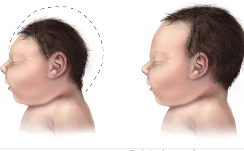
A new study led by researchers at Baylor College of Medicine revealed how in utero Zika virus infection can lead to microcephaly in newborns. The team discovered that the Zika virus protein NS4A disrupts brain growth by hijacking a pathway that regulates the generation of new neurons. The findings point at the possibility of developing therapeutic strategies to prevent microcephaly linked to Zika virus infection. The study appears today in the journal Developmental Cell.
Patients with rare genetic mutations shed light on how Zika virus causes microcephaly
“The current study was initiated when a patient presented with a small brain size at birth and severe abnormalities in brain structures at the Baylor Hopkins Center for Mendelian Genomics (CMG), a center directed by Dr. Jim Lupski, professor of pediatrics, molecular and human genetics at Baylor College of Medicine and attending physician at Texas Children’s Hospital,” said Dr. Hugo J. Bellen, professor at Baylor, investigator at the Howard Hughes Medical Institute and Jan and Dan Duncan Neurological Research Institute at Texas Children’s Hospital.
This patient and others in a cohort at CMG had not been infected by Zika virus in utero. They had a genetic defect that caused microcephaly. CMG scientists determined that the ANKLE2 gene was associated with the condition. Interestingly, a few years back the Bellen lab had discovered in the fruit fly model that ANKLE2 gene was associated with neurodevelopmental disorders. Knowing that Zika virus infection in utero can cause microcephaly in newborns, the team explored the possibility that Zika virus was mediating its effects in the brain via ANKLE2.
In a subsequent fruit fly study, the researchers demonstrated that overexpression of Zika protein NS4A causes microcephaly in the flies by inhibiting the function of ANKLE2, a cell cycle regulator that acts by suppressing the activity of VRK1 protein.
Since very little is known about the role of ANKLE2 or VRK1 in brain development, Bellen and his colleagues applied a multidisciplinary approach to tease apart the exact mechanism underlying ANKLE2-associated microcephaly.
The fruit fly helps clarify the mystery
The team found that fruit fly larvae with mutations in ANKLE2 gene had small brains with dramatically fewer neuroblasts – brain cell precursors – and could not survive into adulthood. Experimental expression of the normal human version of ANKLE2 gene in mutant larvae restored all the defects, establishing the loss of Ankle2 function as the underlying cause.
“To understand why ANKLE2 mutants have fewer neuroblasts and significantly smaller brains, we probed deeper into asymmetric cell divisions, a fundamental process that produces and maintains neuroblasts, also called neural stem cells, in the developing brains of flies and humans,” said first author Dr. Nichole Link, postdoctoral associate in the Bellen lab.
Asymmetric cell division is an exquisitely regulated process by which neuroblasts produce two different cell types. One is a copy of the neuroblast and the other is a cell programmed to become a different type of cell, such as a neuron or glia.
Proper asymmetric distribution and division of these cells is crucial to normal brain development, as they need to generate a correct number of neurons, produce diverse neuronal lineages and replenish the pool of neuroblasts for further rounds of division.
“When flies had reduced levels of Ankle2, key proteins, such as Par complex proteins and Miranda, were misplaced in the neuroblasts of Ankle2 larvae. Moreover, live imaging analysis of these neuroblasts showed many obvious signs of defective or incomplete cell divisions. These observations indicated that Ankle2 is a critical regulator of asymmetric cell divisions,” said Link.
Further analyses revealed more details about how Ankle2 regulates asymmetric neuroblast division. They found that Ankle2 protein interacts with VRK1 kinases, and that Ankle2 mutants alter this interaction in ways that disrupt asymmetric cell division.
The Zika connection
“Linking our findings to Zika virus-associated microcephaly, we found that expressing Zika virus protein NS4A in flies caused microcephaly by hijacking the Ankle2/VRK1 regulation of asymmetric neuroblast divisions. This offers an explanation to why the severe microcephaly observed in patients with defects in the ANKLE2 and VRK1 genes is strikingly similar to that of infants with in utero Zika virus infection,” Link said.
“For decades, researchers have been unsuccessful in finding experimental evidence between defects in asymmetric cell divisions and microcephaly in vertebrate models. The current work makes a giant leap in that direction and provides strong evidence that links a single evolutionarily conserved Ankle2/VRK1 pathway as a regulator of asymmetric division of neuroblasts and microcephaly,” Bellen said.
“Moreover, it shows that irrespective of the nature of the initial triggering event, whether it is a Zika virus infection or congenital mutations, the microcephaly converges on the disruption of Ankle2 and VRK1, making them promising drug targets.”
Another important takeaway from this work is that studying a rare disorder (which refers to those resulting from rare disease-causing variations in ANKLE2 or VRK1 genes) originally observed in a single patient can lead to valuable mechanistic insights and open up exciting therapeutic possibilities to solve common human genetic disorders and viral infections.
BAYLOR COLLEGE OF MEDICINE


