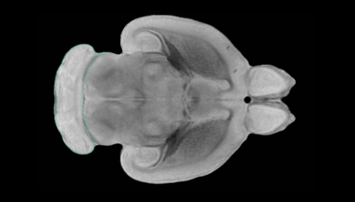
A trio of researchers at Stanford University has found the pathways in the parts of the mouse brain that become active when witnessing pain, fear and pain relief in other mice. In their paper published in the journal Science, Monique Smith, Naoyuki Asada and Robert Malenka describe their study of the neural pathways that are activated in the mouse anterior cingulate cortex (ACC) during empathy and what they learned about them. Alexandra Klein and Nadine Gogolla with the Max Planck Institute of Neurobiology have published a Perspectives piece in the same journal issue outlining the work done by the team in Germany.
Prior research has shown that many animals experience some form of empathy for their peers. As an example, if a dog sees another dog in pain, it will sometimes behave as if it is in pain, as well. Likewise, if a mouse is expressing signs of fear, other mice in the vicinity will almost always begin expressing similar behavior. Humans also experience empathy, of course. However, the brain mechanism responsible for empathy has remained mysterious. In this new effort, the researchers sought to learn more about how empathy works in the mammal brain in general. To that end, they subjected test mice to pain, fear-inducing activities and pain relief while monitoring the brains of observer mice.
Prior work has shown that the ACC is the brain region that is most activated when an animal is experiencing empathy—but the neural circuitry involved has not been studied until now. In order for the brain to process information and then respond to it, parts of the brain are activated as information processing takes place. Such activations are usually followed by electrical signals traveling along neural pathways to other parts of the brain that take part in a response.
To study empathetic response dynamics in the mouse brain, the researchers started by wiring up observer mice to watch their brains in action. Then, observer mice watched as another mouse was shocked, inducing a pain response—they also watched as other mice were scared and as they experienced reductions in pain (though the use of morphine).
The researchers found that as the mice were watching a peer experience pain, the ACC transmitted signals to the nucleus accumbens. When observing a peer experiencing fear, a signal was sent to the basolateral amygdala. Interestingly, they found that signals were also sent to the nucleus accumbens when the observer watched a peer relieved of pain.
Bob Yirka , Medical Xpress

