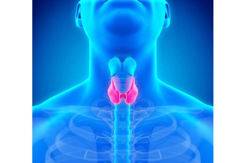
Vocal fold (VF) paresis caused by recurrent laryngeal nerve (RLN) injury is a feared complication of thyroid surgery. It often inflicts a lifelong handicap on the patient; examples are a weak, breathy, and hoarse voice; aspiration problems; and airway distress. In addition to thyroid surgery, all surgical operations on the anatomical path of RLN pose a risk of VF paresis. Iatrogenic VF paresis usually causes symptoms, but it may also be asymptomatic directly after surgery. However early diagnosis of paresis is important; surgeons can obtain immediate feedback, and patients can receive adequate follow-up and treatment. Therefore, after thyroid surgery, screening for VF paresis by laryngoscopy is recommended.
The doctoral dissertation of Maria Heikkinen, Lic Med, inspects the incidence of VF paresis related to different surgical procedures and identifies risk factors for VF paresis in thyroid and parathyroid surgery. The study also investigates the possibility to screen for VF paresis after surgery, with voice assessments comprising perceptual voice analysis by a clinician using the GRBAS (Grade, Roughness, Breathiness, Asthenia, and Strain) scale; patient self-assessment of voice using the Voice Handicap Index (VHI) questionnaire; and acoustic voice analysis using the Multi-Dimensional Voice Program (MDVP).
According to the results, the most common cause for an iatrogenic RLN injury was thyroid surgery (44 percent of all recorded injuries). In contrast, the highest incidence rate for VF paresis was in esophagus surgery (17 percent rate). After thyroidectomy, new postoperative VF paresis was found in 53 of 920 operations (5.8 percent) and in 55 of 1296 nerves at risk of damage (4.2 percent). In hemithyroidectomy, one nerve is at risk; in total thyroidectomy, two nerves are at risk. Furthermore, after 12 months, 55 percent of these paresis cases were uncured. The main risk factors for VF paresis in thyroid surgery were recurrent goiter (odds ratio 5.52, 95 percent CI 2.47-12.30) and malignant thyroid nodule (preoperatively fine needle aspiration biopsy-verified malignancy: odds ratio 4.79 and 95 percent CI 2.08-11.03, malignant final histology: odds ratio 2.61 and 95 percent CI 1.45-4.70).
The GRBAS scale as a screening tool achieved 93 percent sensitivity but only 50 percent specificity in detecting VF paresis after thyroid or parathyroid surgery prior to hospital discharge. The VHI questionnaire had good specificity (90 percent) but poor sensitivity (55 percent). MDVP acoustic voice analysis after surgery prior to hospital discharge or two weeks after surgery achieved only 55 percent sensitivity in identifying VF paresis. None of the voice assessments investigated achieved both adequate specificity and adequate sensitivity to screen for VF paresis after surgery. Combining two measures, one with good sensitivity and one with good specificity, failed to yield a better tool to screen for VF paresis.
In conclusion, thyroid surgery is still the most common surgical procedure causing VF paresis, and the risk of VF paresis in thyroid surgery is the highest among patients with recurrent goiter or thyroid malignancy. The screening voice analysis methods investigated to find VF paresis failed to prove useful, and postoperative laryngoscopy after surgery is still warranted as the most accurate diagnostic test for VF paresis.
The doctoral dissertation of Maria Heikkinen, licentiate of medicine, titled “Vocal fold paresis as a surgical complication: incidence, risk factors, and diagnostic voice analysis” will be examined at the Faculty of Health Sciences.
University of Eastern Finland

