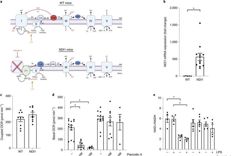
Northwestern Medicine investigators recently discovered that the mitochondrial respiratory chain—a series of protein complexes essential for a cellular respiration and energy production—is necessary for the activation of another protein complex linked to inflammation and the progression of chronic diseases, according to a study published in Nature Immunology.
“Our study revealed that inhibition of mitochondrial respiratory chain dampened NLRP3 inflammasome activation genetically and pharmacologically,” said Navdeep Chandel, Ph.D., the David W. Cugell, MD, Professor of Medicine in the Division of Pulmonary and Critical Care and of Biochemistry and Molecular Genetics, and senior author of the study.
The NLRP3 inflammasome is a protein complex that is activated by viral or bacterial infections and can also cause tissue damage and the progression of chronic disease, including obesity, Alzheimer’s disease and type 2 diabetes, by triggering the release of proinflammatory cytokines.
Previous work had demonstrated that the NLRP3 inflammasome is regulated by mitochondria, specifically through the generation of mitochondrial reactive oxygen species (ROS), notably superoxide and hydrogen peroxide.
In the current study, Chandel and his team used genetic sequencing and pharmacologic strategies to study macrophages (specialized white blood cells) and examine the necessity of mitochondrial ROS for the activation of the NLRP3 inflammasome.
To their surprise, they discovered that mitochondrial ROS were not necessary. Furthermore, inhibitors of the mitochondrial respiratory chain complexes I, II, III and V—all of which are essential for proper production of energy within the cell—prevented NLRP3 inflammasome activation.
They also found that creatine, which is naturally produced by the body and is a widely available nutritional supplement, boosts levels of phosphocreatine, which is essential for proper energy production within cells, and is depleted by the respiratory chain inhibitors, which in turn also sustains activation of the inflammasome.
“The discovery that creatine and phosphocreatine are linked to inflammation opens a new avenue of research with possible implications for creatine,” Chandel said.
According to the authors, the current findings highlight that the mitochondrial respiratory chain sustains NLRP3 inflammasome activation through phosphocreatine-dependent generation of energy and is completely independent of mitochondrial ROS.
Chandel said his team is now interested in investigating how mitochondria control inflammation in the contexts of influenza and Alzheimer’s disease.
“The tools and conceptual framework we generated in this paper will allow us and others to examine how mitochondrial regulate variety of cytokines in distinct diseases,” Chandel said.
Melissa Rohman, Northwestern University

