Astrocytes are star-shaped cells in the brain that play an important role in maintaining the blood-brain barrier, supplying nerve cells with nutrients, and removing metabolic products. At more than 50 percent, they make up the majority of glial cells, the supporting cells in the brain, which until recently were viewed as little more than a kind of “glue” holding nerve cells together. But this view has changed dramatically in recent years, especially for astrocytes.
Astrocytes are now believed to play an important role in signal transduction in the brain through their radiating offshoots (known as astrocyte tendrils), with which they mediate contact between nerve cells and blood vessels. Among other important building blocks, the protein Ezrin is found abundantly in astrocyte tendrils. This suggests that Ezrin is also important for astrocyte function during neuronal development of the brain. Although Ezrin has been studied extensively in vitro in cell culture, up until now there have been few in vivo studies on the role of this protein in astrocytes.
In a recent study, the “Nerve Regeneration” research group led by Prof. Helen Morrison of the Leibniz Institute on Aging—Fritz Lipmann Institute (FLI) in Jena, Germany, has now discovered the role Ezrin plays in brain development and how its absence can prepare the brain to minimize damage after stress, such as a stroke. The study recently appeared in Glia.
What role does Ezrin play in brain development?
“As we know from our own research, in the developing brain, Ezrin is found primarily in developing neurons and is also found in the adult brain in the peripheral protrusions of astrocytes,” reports Prof. Morrison. “But until now, there have been no comprehensive in vivo studies of its functional importance to the nervous system.”
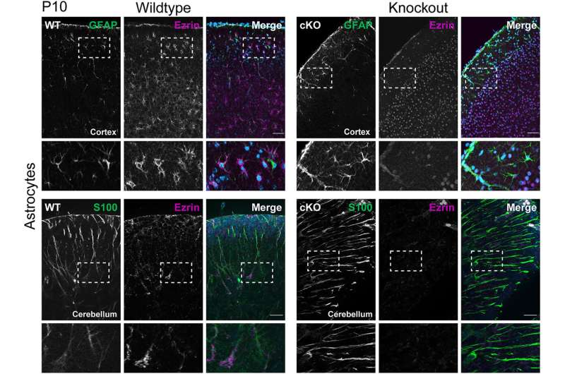
As part of a doctoral research project, in vivo studies were therefore carried out on mice that lacked the protein Ezrin in the nervous system. The investigations focused primarily on the cerebral cortex. Particular attention was paid to the astrocytes and their tendrils in order to investigate in greater detail the role of Ezrin in brain development and in adult brain function.
Ezrin deficiency does not affect brain development
“We were initially quite surprised that the mice developed completely normally despite the lack of Ezrin. Compared to the wild-type mice, they also showed no obvious deficits in learning or memory,” reports Dr. Stephan Schacke, who wrote his doctoral thesis on this topic.
“By all appearances, as the brain develops, structurally and functionally related proteins very similar to Ezrin take over its missing function and thus counteract its loss.” Only when exploring new environments did the modified mice show different, slowed behavior, suggesting changes in neuronal signal processing.
Ezrin deficiency affects glutamate metabolism and alters the shape of astrocytes
Histological methods and proteome analyses were able to demonstrate that the lack of Ezrin alters important cell biological processes, such as glutamate metabolism. Glutamate is one of the most important excitatory neurotransmitters in the central nervous system and is vital for signal transmission between nerve cells.
The strength of signal transmission is controlled, among other things, by the amount of glutamate released and by the length of time until the neurotransmitter is reabsorbed (and thus transmission ends). In signal transmission, an important role is played by the protein GLAST, which is directly involved in glutamate reuptake. In the absence of Ezrin, the amount of GLAST increases, which presumably accelerates the reuptake of glutamate. As a result, signal transmission is both weakened and shortened. This could be a possible explanation for the slower exploratory behavior of the modified mice.
The lack of Ezrin also leads to the upregulation of GFAP, a glial filament protein also found in astrocytes and responsible for their mechanical properties, motility, and cell shape. Increased levels of GFAP are an indicator that astrocytes have undergone morphological changes in their appearance and adopted a “reactive status,” which is also seen in brain damage or disease.
Can the loss of Ezrin help to prevent strokes?
In subsequent studies, it was shown that—compared to wild type mice—the changes in astrocytes triggered by the absence of Ezrin better protect these mice from stress, such as ischemic stroke, in which the brain is no longer supplied with sufficient blood and oxygen due to a blocked artery.
“These mice can withstand a stroke much better than their wild-type relatives because, due to the upregulation of GLAST, they have already learned to mitigate the harmfulness and toxicity of neurotransmitters, especially glutamate, which can lead to stimulus overload and neuronal death if the dose is too high,” explains Prof. Morrison.
“Our study thus not only provides the first important insights into the importance of the Ezrin protein for astrocyte function in our body, but also points to a possible way to achieve improved therapeutic outcome after a stroke if neuronal excitotoxicity—the injury and death of neurons induced by excessive glutamate accumulation—can be efficiently prevented.” Further research will explore this possibility.
Leibniz Institute on Aging – Fritz Lipmann Institute (FLI)

